thyroid nodule thyroid cancer ultrasound colors
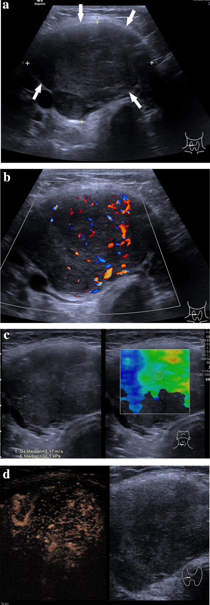
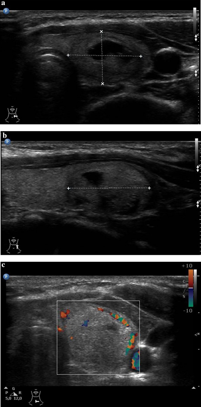
This category of thyroid nodules also has a low risk for malignancy. Pediatric thyroid nodules are often asymptomatic.

Vascularity Assessment Of Thyroid Nodules By Quantitative Color Doppler Ultrasound Ultrasound In Medicine And Biology
Vascular fraction area mean flow velocity index and flow volume index in three regions nodule cente.

. This Nodule Shown In Red Comprises About 80 Of The Thyroid Tissue Shown In Yellow In This Particular Area Of The Thyroid. Keep in mind however that an ultrasound alone cannot make the. We searched the PubMed Web of Science Embase and.
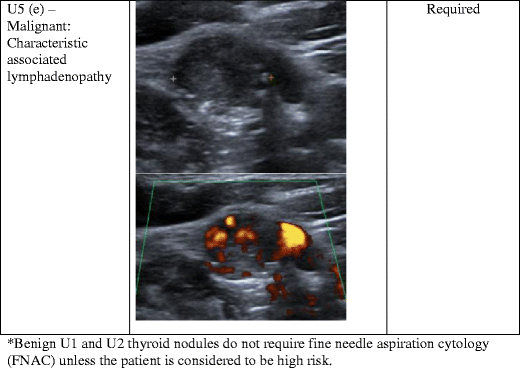
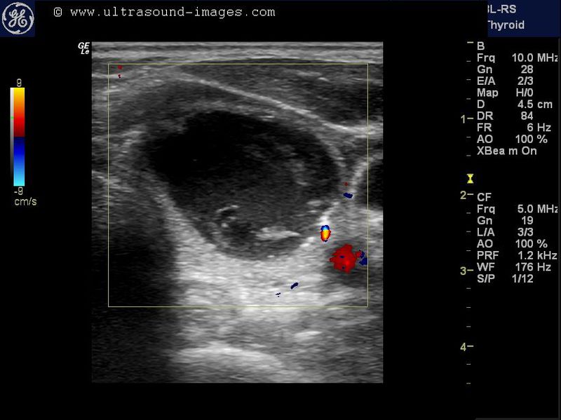
This study aimed to analyze the value of color Doppler ultrasound in the diagnosis of thyroid nodules. Vessels in which blood is flowing are colored red for flow in one direction and blue for flow in the other with a color scale that reflects the speed of the flowbecause. Automatic segmentation of the thyroid.
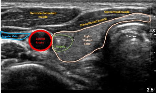
Ultrasound features related to thyroid lesions structure shape volume and margins are considered to determine cancer risk. Thyroid Nodule Thyroid Cancer Ultrasound Colors. The probe is placed on the skin which is at the very top of the picture and sound waves are directed deep into the neck and thyroid.
Color Doppler images of 100 nodules were analyzed for three metrics. This study suggests that ultrasound features of microcalcifications solid nodule and size larger than 2 cm can be used to identify patients at high risk for thyroid cancer. Ultrasound elastography USE proved to be a valuable imaging tool in predicting the risk of malignancy of thyroid nodules 20 21 and also in decreasing the FNAC indication.
Nodules that are found to be malignant have with a higher likelihood of locoregional metastasis to the lateral neck or distant. The outcome indicators in the articles had to include the numbers of true. Ultrasound uses soundwaves to create a picture of the structure of the thyroid gland and.
In this group both the thyroid nodule and the surrounding thyroid tissue have green appearance. The Doctor Uses A Very Thin Needle To. This study aimed to analyze the value of color Doppler ultrasound in the diagnosis of thyroid nodules.
Certain characteristics of thyroid nodules seen on an ultrasound are more worrisome than others. Benign thyroid nodule On US thyroid nodules are depicted as discrete lesions as they cause distortion of the homogeneous echo pattern of the. A common imaging test used to evaluate the structure of the thyroid gland.
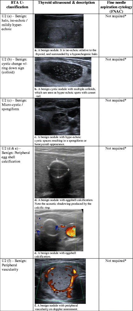
The only way to definitively determine if a thyroid nodule is cancerous is to examine it under a microscope. The most common method is called fine need aspiration FNA. Ultrasound classification U2.
Using Artificial Intelligence To Predict Risk Of Thyroid Cancer On Ultrasound Imaging Technology News

Pdf Ultrasonographic And Color Doppler Ultrasonographic Parameters To Discriminate Thyroid Nodules Semantic Scholar

Us Features Of Thyroid Malignancy Pearls And Pitfalls Radiographics

Us Features Of Thyroid Malignancy Pearls And Pitfalls Radiographics

Thyroid Nodule Ultrasound What Is It What Does It Tell Me

Ultrasonographic Features For Differentiating Follicular Thyroid Carcinoma And Follicular Adenoma Sciencedirect

Incidental Finding Of An 18fdg Positive Thyroid Nodule On Pet Ct Imaging

Characterization Of Malignant Solid Thyroid Nodules By Ultrasound And Doppler Semantic Scholar
5 Best Ways To Diagnose Thyroid Cancer
Pattern Recognition Of Benign And Malignant Thyroid Nodules Ultrasound Characteristics And Ultrasound Guided Fine Needle Aspiration Of Thyroid Nodules Radiology Key
Ultrasonography Of Thyroid Nodules A Pictorial Review Insights Into Imaging Full Text
Ultrasound Findings Of The Thyroid Gland In Children And Adolescents Springerlink
A Gallery Of High Resolution Ultrasound Color Doppler 3d Images Thyroid 2
Ultrasound Findings Of The Thyroid Gland In Children And Adolescents Springerlink
Ultrasonography Of Thyroid Nodules A Pictorial Review Insights Into Imaging Full Text

Conventional Ultrasound Of Papillary Thyroid Carcinoma Using Aa And Download Scientific Diagram

Sonographic Features Suggestive Of Papillary Thyroid Carcinoma
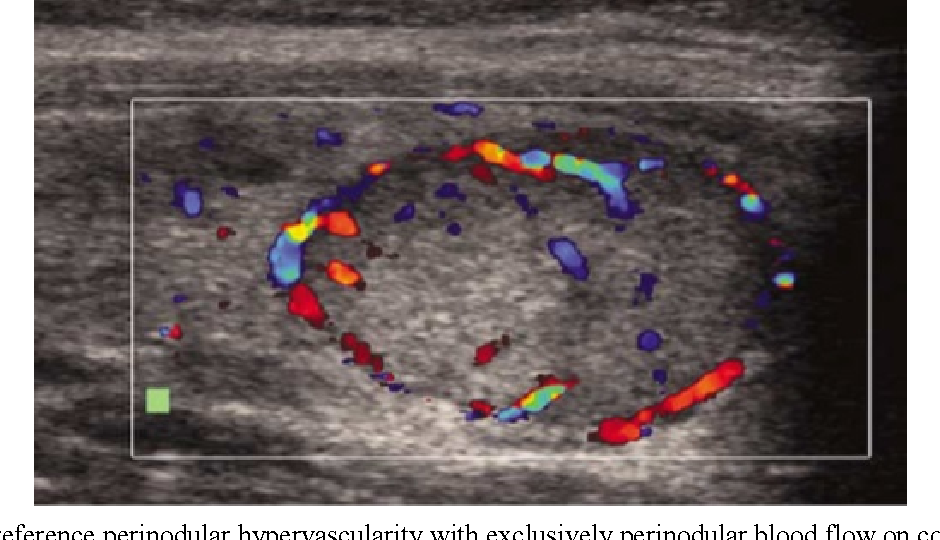
Figure 3 From Gray Scale Vs Color Doppler Ultrasound In Cold Thyroid Nodules Semantic Scholar
